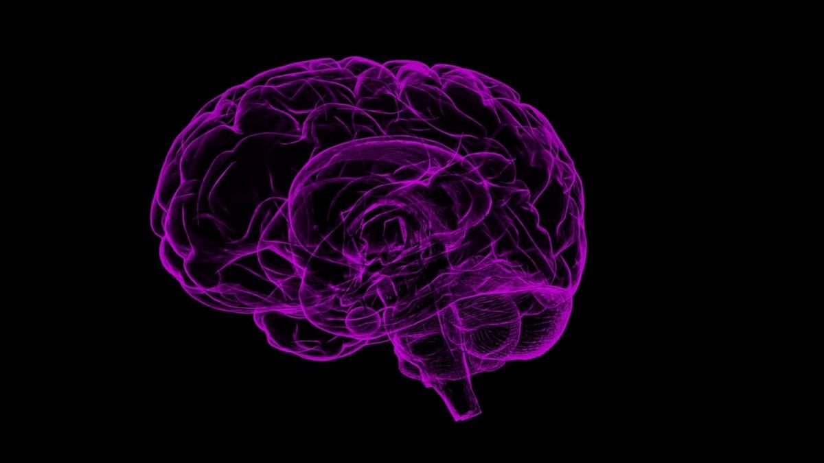
Captivating activity lights up a lab-grown model that acts more like a living network than a static sample. Built to link regions that rarely communicate in a dish, it offers a fresh path to study whole-brain disorders without revealing more than the title allows. In clear terms, this breakthrough mini human brain brings coordinated signals, early barriers, and measurable structure into view, while keeping the story focused, human, and easy to read.
A whole-brain model that finally acts in concert
Researchers assembled a small organoid that includes tissues from multiple brain regions working together. Instead of isolating a cortex or a hindbrain, they fused parts so they could interact and spark network responses. The result behaves like a coordinated system, not five separate islands, which changes how scientists can observe development.
They verified the fusion with light-sheet microscopy, which captures glowing signals across layers without damage. That view confirmed that separate organoids merged into one structure and produced electrical activity. The signals did not fire at random. They formed patterns that suggest region-to-region dialogue, which is key for studying complex conditions.
An early blood-brain barrier also began to form. That layer controls what can cross into brain tissue and matters for drug testing. Seeing it appear inside a small model helps align experiments with human biology. It also gives teams a way to measure permeability while networks mature.
What this mini human brain is made of
The team first grew several pieces in separate dishes: cerebral organoids for brain tissue, endothelial organoids for rudimentary vessels, and midbrain or hindbrain components. After that step, they guided the parts to meet. Proteins that behave like biological glue helped them stick long enough to connect and grow.
As the fused structure matured, it produced electrical signals that echoed network behavior. Those signals hinted at circuits forming across regions. Instead of single-area spikes, the activity showed coordination. That shift matters, because whole-brain disorders rarely sit in one location. They ripple through systems and timelines.
Compared with single-region organoids, the multi-region build adds context. It combines tissues that typically talk in the body. When regions interact, scientists can watch cause and effect that would be missed in isolation. That broader view supports stronger hypotheses and fewer experimental dead ends.
From dish to insight: uses that matter
A human cell-based model lets teams follow neurodevelopment as it unfolds. With signals tracked in real time, researchers can observe when connections appear strong, weak, or mistimed. Because timing errors often drive symptoms, this moving picture is more useful than snapshots from fixed samples.
The organoid retained diverse neuronal types similar to a 40-day-old human fetus. About 80% of early cell types appeared at comparable levels, which keeps experiments grounded in human biology. Though small, it held around 6 to 7 million neurons, enough to model early patterns while staying practical for lab work.
This platform targets brain-wide conditions such as autism, schizophrenia, and Alzheimer’s. It can show how a pathway goes off track and when that shift begins. With a single mini human brain, teams can compare patterns before and after an intervention, then decide which signals deserve deeper study.
Why a mini human brain could speed safer drugs
Drug programs fail too often, especially for brain conditions. Roughly 85% to 90% of candidates do not clear Phase 1. For neuropsychiatric drugs, failure rates near 96% raise costs and delay relief. Better models may improve those odds by filtering weak ideas earlier.
Whole-brain organoids more closely mirror human development than animals do. When one model contains interacting regions and an emerging barrier, responses look more realistic. That relevance helps teams rank targets, select doses, and drop risky compounds before trials, which saves time and protects participants.
Because the structure uses human cells, it can also support personalized testing. Teams can compare patient-specific organoids and see which therapy shifts a network toward typical patterns. When a signal improves inside the dish, it offers a data-driven reason to advance a candidate or combine treatments.
Limits, safeguards, and the road to translation
The organoid is not a full brain. It lacks the size, maturity, and tens of billions of neurons seen in adults. Even so, the 6 to 7 million neurons inside create enough complexity to study early activity. That balance of scale and control is what makes the platform powerful.
Ethical care remains central. The model shows coordinated activity, not consciousness. It glows because of imaging and markers, not self-aware signals. Light-sheet microscopy provides vivid views while keeping tissue intact, so scientists can track growth without pushing the system beyond intended limits.
Next steps focus on perfusion, stability, and richer readouts. Adding better flow may support longer studies. Aligning signals with behavior in animals can bridge the lab to the clinic. Each improved mini human brain should refine targets, sharpen timelines, and help translate basic insight into actionable care.
What today’s result means for tomorrow’s brain research
Published in Advanced Science, this multi-region model shows that fused organoids can talk, build early barriers, and light up as connected networks. Those features offer a rare mix of depth and practicality. With human biology, clearer signals, and space to test therapies, the path from hypothesis to help looks closer.